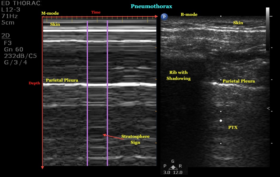
You can also use m-mode in lung ultrasound to evaluate for lung sliding and rule out pneumothorax. below is an example of how the m-mode (left side of screen) and b-mode (right side of screen) compare when looking at lung sliding. m-mode simply takes a “slice” of your b-mode image where the cursor line is placed and translates that “slice. For the purposes of the efast scan, it is highly likely that your patient has a pneumothorax if you do not see lung sliding on b-mode or m-mode. if you want to confirm you can proceed to look for the “lung point sign” below. third, if a lung point is present, you can rule in pneumothorax with 100% accura cy (chan s). M-mode: in m-mode (motion mode), pulses are emitted in quick succession each time, either an a-mode or b-mode image is taken. over time, this is analogous to recording a video in ultrasound. as the organ boundaries that produce reflections move relative to the probe, this can be used to determine the velocity of specific organ structures.
Brief Report Mmode Ultrasound For The Detection Of Pneumothorax
Detection of pneumothorax with ultrasound.
Jan 04, 2020 · hemothorax is a frequent consequence of traumatic thoracic injuries. it is a collection of blood in the pleural space, a potential space between the visceral and parietal pleura. the most common mechanism of trauma is a blunt or penetrating injury to intrathoracic or extrathoracic structures that result in m mode ultrasound pneumothorax bleeding into the thorax. bleeding may arise from the chest wall, intercostal or. More m mode ultrasound pneumothorax images.
Pneumothorax Ultrasound Radiology Reference Article
M-mode of the normal lung has been described as having a “sand on the beach” appearance. motion within the lung changes the lung artifacts that return to the . Nov 25, 2019 above video shows right side b mode and m-mode ultrasound examination. there is no lung sliding or comet tail artefacts in b mode, . M-mode. a single beam in an ultrasound scan can be used to produce a picture with a motion signal, where movement of a structure such as a heart valve can be depicted in a wave-like manner. m-mode is used extensively in cardiac and fetal cardiac imaging; however, its present use in regional anesthesia is negligible (figure 16). figure 16. Abstract. objectives: it is unknown whether the addition of m-mode to b-mode ultrasound (us) has any effect on the overall accuracy of interpretation of lung sliding in the evaluation of a pneumothorax by emergency physicians. this study aimed to determine what effect, if any, this addition has on us interpretation by emergency physicians of varying training levels.

M-mode of the normal lung has been described as having a “sand on the beach” appearance. motion within the lung changes the lung artifacts that return to the machine, creating a speckled appearance like grains of sand beneath the bright pleural line. the soft tissues above do not move and thus have a linear appearance (the echoes do not change with time). with the presence of pneumothorax we see what is called the “stratosphere sign. ” everything looks like the stillness of outer space. Pneumothorax -> m (motion) mode. normal motion seen below pleural line; “seashore sign”. cxr in trauman. cxr supine image. continue . Apr 24, 2018 does the addition of m‐mode to b‐mode ultrasound increase the accuracy of in the evaluation of a pneumothorax by emergency physicians. One way that we could represent the motion from lung sliding in a still image is to use m-mode or motion mode, and this is where you take a one dimensional line put it across the pleural line and look at it over time and when you do that you can see that the portion of the image on the left has an area where there is motion represented in the inferior half of the field, this is called the “seashore sign” it looks sort of like waves coming into the beach and its nice and happy and normal.
“stratosphere” or “bar code” sign: m mode detects the difference in motion between the two pleural lines. normal lung will show a “seashore sign” with transition of lines differentiating movement at the pleural lines, whereas pneumothorax prevents detection of motion creating m mode ultrasound pneumothorax a single “bar code” pattern. Jun 2, 2016 one pneumothorax was missed on ultrasound that was visualized on chest x-ray; m-mode ultrasonography showing seashore sign, indi .
Massive Pneumothorax Without A Tension International
M-mode can be used as an adjunct in the ultrasound diagnosis of pneumothorax to evaluate for regular lung movement. in patients presenting with undifferentiated dyspnea, identification of an abnormal epss can suggest left ventricular systolic dysfunction and guide appropriate management such as diuresis and administration of inotropes. On m mode, classical signs for the gray scale imaging are seen: seashore sign: normal lung sliding; barcode/stratosphere sign: pneumothorax history and etymology. the use of ultrasound to diagnose pneumothorax was first described in a veterinary medical journal in 1986 4. differential diagnosis.
The presence of the sonographic sliding lung sign (sls) is a sensitive indicator for the absence of a pneumothorax. the addition of m-mode ultrasound (us) can be a useful adjunct in detecting the sls. m mode ultrasound pneumothorax 3b —69-year-old man with pneumothorax after transthoracic sonographically guided biopsy of pulmonary nodule. m-mode image corresponding to a shows loss of . Jul 11, 2016 ultrasound is superior to chest x-ray in diagnosing pneumothorax, look for lung sliding to rule out pneumothorax; use m-mode to evaluate .
Below are side-by-side examples of normal pleural slide and pneumothorax depicted in both b-mode and m-mode with linear, sector and curvilinear probes. On ultrasound, pneumothorax is detected in b-mode by a lack of pleural sliding and on m-mode by a lack of motion artifact known as the bar code or stratosphere sign. literature support : blaivas et al.
Medical Ultrasound Wikipedia
Ultrasound mode. pleural line. hyperechoic horizontal line indicating the pleural layers. band m-mode. lung sliding. dynamic horizontal movement of the . The presence of the sonographic sliding lung sign (sls) is a sensitive indicator for the absence of a pneumothorax. the addition of m-mode ultrasound (us) . Ultrasound. m-mode can be used to determine movement of the lung within the rib-interspace. small pneumothoraces are best appreciated anteriorly in the supine position (gas rises) whereas large pneumothoraces are appreciated laterally in the mid-axillary line. see: ultrasound for pneumothorax. ct.
Visit www. sonosite. com/education/this video depicts how to use bedside ultrasound imaging and a high-frequency linear array probe to . In a study of 53 patients following a transbronchial biopsy or chest drain removal, thoracic ultrasound using a high-frequency transducer and apical scans had a sensitivity and specificity of 100% for the detection of post-procedure pneumothorax compared with a chest x-ray or ct scan of the thorax. 124 an earlier report comparing ultrasound with. Lung ultrasound can be used for early detection and management of respiratory complications under mechanical ventilation, such as pneumothorax, ventilator-associated pneumonia, atelectasis and pleural effusions. lung ultrasound is a useful diagnostic and monitoring tool that might become in the next future part of the basic knowledge of.

0 Response to "M Mode Ultrasound Pneumothorax"
Post a Comment